ENT Health

Expert Ear, Nose and Throat News – Now!

Saturday, December 15, 2012

Voice Amplification for Patients with a Weak Voice

No comments

There are certain voicebox conditions that naturally tends to a weak sounding voice.

For some, the voice is weak all the time sounding breathy or just lacks projection. For others, the voice starts out OK, but than becomes weak after talking for some period of time. There are both pathological (ie, vocal cord paralysis) as well as functional (ie, muscle tension dysphonia) causes of a weak voice, but in the end, a patient just wants a better sounding voice one way or another.

Although voice therapy is a standard way of addressing vocal weakness regardless of causation as well as possible surgical interventions, patients still need to use their voice and in order to obtain an immediately improved vocal strength is to use electronic amplification.

For the same reason a pop star singer uses a microphone to amplify their voice to be heard in a gigantic stadium, a person with a weak voice can use a similar system to amplify their voice in more mundane situations.

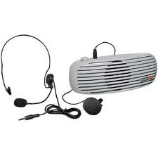
In essence, such personal voice amplification systems contain 2 components: microphone and speaker. The microphone is placed near the mouth and the speaker placed near the person, typically on a belt.
Such systems are relatively cheap costing less than $100. Many models can be found on Amazon.comat a discount.

The "premium" voice amplification systems are by Chattervoxand cost several hundred dollars.

Some examples can be found here.


Thursday, December 13, 2012

Julie Andrews and Her "Botched Throat Surgery"

No comments

It has already been well-publicized that the former singing sensation Julie Andrews underwent some type of throat surgery in 1997 after which she never regained her phenomenal 4-octave voice thus relegating her to more conventional roles of being an actress without the singing. This "botched throat surgery" per Julie Andrews lead to a lawsuit that was ultimately settled.

It is unknown precisely what type of (presumed) vocal cord pathology was present as well as what type of surgical approach was used to try and fix it. In a recent interview, she stated:
"The operation that I had left me without a voice and without a certain piece of my vocal chords"
However, given her aggressive and active singing career prior to surgery, she probably had vocal cord nodules, a benign growth that occurs due to vocal overuse, a situation not uncommon with professional singers.

Vocal cord nodules are most always due to excessive voice use leading to "callous" formation on the vocal cord lining, much like shoveling dirt will eventually lead to callous formation on the hands.

Traditional standard of care management of vocal cord nodules is voice therapy and avoidance of any activities leading to voice abuse (screaming, yelling, etc). However, resolution of nodules with such behavior focused treatment takes months. However, although it takes a while for the nodules to resolve with this treatment method, they typically do not come back.

For patients who are more "impatient" for results (not unusual with professional singers whose livelihoods depend on singing), there are more aggressive ways to address vocal cord nodules with possible resolution within weeks. However, the caveat is if the underlying voice behavior that led to nodule formation in first place is not addressed, the vocal cord nodule WILL recur after initial resolution/improvement. Furthermore, as with any more invasive treatments to obtain a "quick fix", scar formation may occur leading to permanent voice changes, usually for the worse which obviously happened with Julie Andrews.

What are some of these more invasive treatments beyond voice therapy? To reiterate, these procedures also have a high risk of recurrence if underlying abusive voice behavior that led to the nodule formation in first place is not first addressed.

• Surgical excision can be performed, but can lead to permanent scar formation during the healing process that can lead to persistent irreversible hoarseness.

• Botox injection can also be pursued which causes a "partial" vocal cord paralysis preventing the repetitive trauma in the region of the vocal cord nodule.

• Steroid injection to the vocal cord nodule(s) can possibly resolve or reduce the nodule resulting in improved vocal quality within weeks. Such local injection technique has mainly been performed in the treatment of spasmodic dysphonia (botox injection), vocal cord granulomas, and vocal cord paralysis. Watch a video how a "local injection" to the vocal cord can be performed (video shows injection of vocal cord granuloma rather than nodule, but overall approach is identical).

Read more about vocal cord nodules here.

Source:
Julie Andrews' Voice Isn't Coming Back, But She's Not Staying Silent. Huffington Post 12/5/12

PUBLIC LIVES; Julie Andrews Sues Throat Surgeon. NYT 12/15/99

Julie Andrews Settles Lawsuit Against Doctors. ABC News 9/7/12

Monday, December 10, 2012

Coffee Reduces Risk of Death from Oral Cancer

No comments


In a prior blog, I mentioned that researchers found that regular coffee drinkers who drank more than four cups of coffee a day had a 39 percent decreased risk of two types of head and neck cancer: oral cavity and pharynx cancers. In the study published in the Journal of Cancer Epidemiology, Biomarkers & Prevention, they found however, that coffee did not decrease the risk of laryngeal cancer. Read a CNN report on this here.

In a more recent study (Dec 2012), researchers have found that not only does coffee reduce risk of cancer, but that consuming more than four cups of caffeinated coffee per day was associated with a 49 percent lower risk of oral/pharyngeal cancer death relative to no/occasional coffee intake.

Coffee contains over 1,000 different chemical compounds, including cancer-fighting antioxidants, and it’s those antioxidants that may provide a “plausible explanation” for reducing the cancer risk as well as cancer death.

However, patients should not take this study as an excuse to increase their coffee intake specifically to counteract known cancer-causing behaviors including smoking, drinking alcohol, and chewing tobacco.

Reference:
Coffee and Tea Intake and Risk of Head and Neck Cancer: Pooled Analysis in the International Head and Neck Cancer Epidemiology Consortium. Cancer Epidemiol Biomarkers Prev. 2010 Jul;19(7):1723-36. doi: 10.1158/1055-9965.EPI-10-0191. Epub 2010 Jun 22.

Coffee, Tea, and Fatal Oral/Pharyngeal Cancer in a Large Prospective US Cohort. American Journal of Epidemiology, 2012 DOI: 10.1093/aje/kws222

Sunday, December 9, 2012

Embedded Earrings and Treatment

2 comments

A few times a month, I have a patient, usually a young child, who presents with an earring that not only can not be removed, but is embedded within the earlobe (or ear) itself.

There are two common scenarios that lead to this unfortunate situation: infection and fiddling.

Infection is fairly straightforward. For whatever reason, the piercing gets infected and the earlobe skin starts to swell around and eventually even enveloping the earring itself. To prevent an embedded earring, immediately remove the earring!!! If you wait until the swelling is too severe, it may not be possible to remove the earring at all without surgical intervention. This type of infection can occur rapidly from being normal to rip-roaring infected swelling within 24 hours.

Fiddling is basically when the patient keeps tightening the earring backing snug with the skin as tightly as possible so the earring won't loosen and potentially get lost. However, such tightening can actually induce the skin to overgrow the earring with subsequent infection. Always make sure there is some “play” that allows the earring to push back and forth through the ear and turn easily.


Now, should the earring become embedded and manual removal not possible, surgical intervention is required.

The steps are as follows:

1) Sterilize the area with betadine.
2) Inject area with lidocaine.
3) After the area becomes anesthetized, a scalpel is used to create an "X" shaped incision over the embedded earring.
4) Toothed forceps and clamps are used to grab and pull the embedded earring out.

Aftercare typically involves oral antibiotics and application of antibiotic ointment several times a day. Once the infection clears, I typically recommend waiting a few months before re-piercing to allow scar activity to completely fade first.

Also, keep in mind that they DO make earrings that are physically impossible to tighten up against the skin to reduce the possibility of embedded earrings.

On that note, we do offer ear-piercing in our office.

Tuesday, November 27, 2012

Is Patient Abandonment in the Operating Room Ever Justified?

No comments

I ask this question mainly because there WAS an ENT who WAS sued (and settled) for NOT abandoning his patient in the operating room. [link]

Imagine this hypothetical scenario...

You are an ENT surgeon in a rural community hospital performing a fairly difficult but elective sinus surgery on a 12 years old child with cystic fibrosis with extensive pan-sinus nasal polypoid disease. Given the extensive nasal polypoid disease, a fair amount of bleeding is occurring which was not unanticipated.

Suddenly, you get a phone call from the emergency room regarding a patient with progressive difficulty breathing suspected to have epiglottitis.

What do you do if you are the only ENT in the hospital?

Do you "abandon" the 12 years old child in the operating room, still bleeding, still under general anesthesia and attend to the ER patient who himself might die without an emergency tracheostomy? Doing an evaluation to determine how critical the ER patient may take anywhere from 15 - 60 minutes including the tracheostomy itself.

What if you are the only ENT available in the region let alone the hospital? No fellow ENT colleagues to call upon for help. General surgery is "unavailable" or not comfortable with performing tracheostomies especially given ENT performs all tracheostomies in the hospital?

Patient abandonment is defined as:
  • Failing to transfer a patient to an appropriate level of care
  • Failing to respond to calls from a hospital regarding a patient
  • Refusing to care for a patient after arranging the patient's admission
  • Failing to treat a patient until new coverage is arranged
Proving patient abandonment includes:
  • Your doctor had a duty to treat you - a duty was created when the physician-patient relationship was established
  • You had a reasonable expectation that your doctor would treat you
  • Your doctor failed to treat you although he or she was obligated to do so
  • You suffered injury as a result
Well, according to one lawsuit, it seems that the ENT was required to abandon his patient in the operating room and attend to the ER patient. The lawsuit stemmed from the fact that the ENT did not abandon his patient in the operating room and the ER patient did die as a result of not being attended to quickly and a tracheostomy performed. Of course, the settled lawsuit also blamed the hospital, general surgery, and anesthesiology.

However, abandoning a patient on the operating room table is also tantamount to medical malpractice according to the very definition of patient abandonment.

And, I would not be surprised if the patient on the operating room table would have sued the ENT if he DID leave the operating room in the middle of surgery to attend to another patient.

What to do?

I have no answer...

Doing an emergency tracheostomy is HARD, even for someone who has performed hundreds of elective tracheostomies. I know... I've done perhaps a half-dozen emergency trachs in my career so far. In this particular lawsuit, I found it incredible that a hospitalist (not a surgeon) was the one who finally attempted the emergency tracheostomy (albeit unsuccessfully).

Do you consider the patient you are CURRENTLY caring for has a higher priority than a patient you have never met, even if possible life-threatening illness is involved? (Keep in mind that when called for an airway problem, that 99% of the time, an emergency tracheostomy is not needed.)

OR, do you prioritize the patient you have never met given the possible life-and-death circumstances involved, even if 99% of the time, no surgical airway is required.

What would YOU do? What should you do? Feel free to comment below!

Source:
Hospital settles wrongful death lawsuit. Curry Coastal Pilot 10/31/12

Saturday, November 17, 2012

Taste Changes after Tonsillectomy

No comments

Very rarely, patients will complain of taste changes after tonsillectomy. Such taste changes (aka dysgeusia) is most often described as metallic though other taste variations or absences may be reported.

Depending on what study you look at, this complaint occurs anywhere from 0.3% to as high as 9% of tonsillectomy cases. Dysgeusia after tonsillectomy is felt to be due to a number of different causes including:

1) medication side effect
2) injury to the lingual branch of the glossopharyngeal nerve
3) zinc deficiency

Regardless of the etiology, most cases of post-tonsillectomy dysgeusia spontaneously resolves within a few months without any specific intervention.

However, there are a few things a patient can try that might help accelerate normal taste recovery including:

1) Stopping all unnecessary medications
2) Zinc gluconate 50mg 3 times per day
3) Alpha Lipoic Acid 200mg 3 times per day
4) Rinsing mouth with watered down milk-of-magnesia
5) Chewing non-mint flavored gum

I did want to point out that there are no comprehensive studies to support any of these interventions. But, it certainly can't hurt to try it.

References:
Taste disturbance after tonsillectomy. Acta Otolaryngol Suppl. 2002;(546):164-72.

Taste disorders after tonsillectomy: case report and literature review. Ann Otol Rhinol Laryngol. 2005 Mar;114(3):233-6.

Posttonsillectomy taste distortion: a significant complication. Laryngoscope. 2004 Jul;114(7):1206-13.

Taste disturbance after tonsillectomy and laryngomicrosurgery. Auris Nasus Larynx. 2005 Dec;32(4):381-6. Epub 2005 Jul 19.

Thursday, November 8, 2012

Brooke Burke Has Thyroid Cancer

No comments

Brooke Burke, TV personality best known for winning the 7th season of Dancing with the Stars and later co-hosting the show starting in 2010 announced in a self-published video the fact that she has thyroid cancer and will soon be undergoing thyroid surgery.

Apparently, on a routine physical exam, a thyroid nodule was appreciated in July 2012. This was biopsied (presumably via an ultrasound guided fine needle aspiration) and came back as "atypical". Given atypical findings, a diagnosis of cancer or no cancer can not be definitively made. As such, more studies were than pursued and finally, she was informed she had a "good" type of thyroid cancer if there is such a thing.

Some of these additional studies may have included CT scan of the neck and thyroid scan.

The type of thyroid cancer was never specifically mentioned, but she most likely has papillary thyroid carcinoma which is the most common form of thyroid cancer and is highly curable (which is where the comment of being a "good" cancer comes from).

Other less common thyroid cancers include follicular thyroid carcinoma and medullary thyroid carcinoma.

Thyroid cancer mandates thyroidectomy with post-operative radioactive iodine treatment to ensure complete thyroid tissue eradication.

Her surgery date has not been announced.

It should be stated that until the thyroid is removed, one can NOT be absolutely sure that thyroid cancer actually truly is present. Indeed, the president of Argentina made a similar announcement of thyroid cancer only to find out after her thyroid was removed that no cancer was actually present. Read blog post about this "mistake".

Source:
Brooke Burke: I Have Cancer. NY Post 11/8/12


Sunday, November 4, 2012

Breathing or Voice... You Can Only Have One...

No comments

Sounds like a choice taken out of a morbid horror movie...

"You can ONLY have one... Your voice or your breath..."

The victim than loses one or the other after making a choice getting either the tongue or head cut off with a knife.

But patients with vocal cord paralysis on both sides are essentially faced with this very question.

Normally, the vocal cords (TVC in picture below) found in the voicebox move to accommodate talking or breathing.


When the vocal cords are apart, breathing occurs allowing air to pass unimpeded between the vocal cords and down into the lungs. However, with talking, the vocal cords come together tightly and vibrate creating voice. Watch a movie.

However, when both vocal cords become paralyzed, they neither move apart to help with breathing nor come together to assist with talking. Indeed, the voice and breathing BOTH stink.

It is in just this situation that the choice of breathing or voice becomes important because a patient can not have both. Improving one will sacrifice the other.

To explain this connection of voice and breathing further, normally, when the vocal cords are both moving fine, the voice is at 100% and breathing is at 100%. However, with bilateral paralysis, the voice and breathing are now linked to each other and collectively can not exceed 100%. As such, a patient with new onset bilateral vocal cord paralysis may start with a 40% of normal vocal quality and 60% of normal breathing ability for a total of 100%.

Using this scenario as a starting point where vocal quality is at 40% and breathing is at 60% of normal...

If a patient chooses to improve the vocal quality from 40% to 100% (an improvement of 60%), then breathing WILL correspondingly decrease 60% down to 0%... and potentially die given the ability to breathe is lost. Why? Because the vocal cords can be surgically forced together to allow for voice... but given the vocal cords are paralyzed, they can not move back apart to allow for breathing.


If a patient chooses to improve breathing quality from 60% to 100% (an improvement of 40%),  then the vocal quality WILL correspondingly decrease 40% down to 0%... and lose the ability to talk. Why? Because the vocal cords can be surgically forced apart to allow for breathing... but given the vocal cords are paralyzed, they can not move back together to allow for vocal cord vibration.


The total percentage of vocal and breathing quality can never be more than 100%. If a patient wants the best possibly voice and breathing, than a compromise would be to increase the vocal quality 10% from 40% to 50%, but understanding that this 10% improvement in vocal quality WILL mean a corresponding 10% decrease in breathing ability from 60% to 50%. This 10% change means that in the end, the voice and breathing would both end up at 50% of normal.

In most cases of bilateral vocal cord paralysis, it is the reduced breathing and shortness of breath that bothers people the most. The voice isn't good, but when given the choice, most people choose to try and improve their breathing ability understanding their voice may further deteriorate.

To read more about such treatment options, click here.

Saturday, November 3, 2012

Nasal Sounding Speech

No comments

There are actually TWO different flavors of nasal sounding speech.

HYPO-nasal speech is due to reduced or no nasal airflow and HYPER-nasal speech due to too much nasal airflow.

HYPO-nasal speech is by far the most common cause of nasal-sounding speech. It is similar to how a person would sound if they pinched their nose shut while talking. As such, ANY anatomic nasal obstruction would by definition lead to such hypo-nasal speech. Treatment, obviously, is to remove this anatomic nasal obstruction whatever it may be either with medications or surgery. Examples of hypo-nasal speech causes include:
HYPER-nasal speech itself has several different flavors, but the key concept is the presence of an opening between the mouth and nose when it should be sealed shut during speech. Normally, complete closure should occur with certain sounds like /s/, /sh/, /b/, and /p/. Such sounds are called plosives and sibilants. This link provides a cartoon animation of how each sound in the English language is produced from an anatomical standpoint.

Now what are some of the causes of HYPER-nasal speech? Causes can be divided broadly into either anatomic and functional variants.

Anatomic HYPER-nasal speech include:
Functional HYPER-nasal speech include:
  • Velopharyngeal insufficiency without anatomic cause. Velopharyngeal insufficiency or VPI occurs when the soft palate does not seal against the back of the mouth during appropriate speech sounds.
  • Poor articulation (person is not correctly pronouncing words)
Treatment of HYPER-nasal speech may include speech therapy and surgery depending on the cause.

Given the subtleties involved in differentiating the different causes of nasal speech, workup typically involves not only just looking in the nose or mouth, but also performing a nasal endoscopic examination.

Below is a video showing a young child undergoing just such an endoscopic exam. Here are some other videos containing audio of abnormal endoscopic exams.

Tuesday, October 30, 2012

Elbowed by Your Spouse While Sleeping? You May Have Sleep Apnea!

1 comment

Canadian researchers have determined that if a patient answers in the affirmative to two questions:

1) Do you get elbowed/poked while sleeping for snoring?
2) Do you get elbowed/poked while sleeping for stopping breathing?

There is a significant chance that the patient may have obstructive sleep apnea (OSA) with AHI score  more than 5.

This quiz has been dubbed the "Elbow Test".

Actual diagnosis for OSA is by sleep study.

Should OSA be actually diagnosed on a sleep study, initial treatment includes CPAP machine followed by oral appliance and potential candidacy for surgical interventions.

Reference:
“Elbow Test” May Predict Sleep Apnea. Chest 10.22.12

Wednesday, October 24, 2012

"Mother's Kiss" to Remove Nasal Foreign Bodies

No comments

In 1965, a general practitioner Vladimir Ctibor described the "Mother's Kiss" technique of removing nasal foreign bodies in a child.

Essentially, a trusted adult (like a mother) places her mouth over the child's mouth as if to perform mouth-to-mouth resuscitation. While pinching off the unaffected nostril, the adult than blows gently into the child's mouth until resistance is felt caused by the child closing the glottis. At that moment, the adult gives a sharp explosive exhalation to deliver a strong puff of air that passes up into the nose and out the unblocked nostril. If successful, this air puff will also blow the foreign body out the nose as well.

If the adult blow's air when the child's glottis is open, air will just go into the lungs rather than up the nose.

Prior to the procedure, the child is informed that the mom will give the child a "big kiss," hence the name of this procedure.

Now... does this technique actually work?

According to one meta-analysis, it works about 59.9% of the time.

The way I consider it... it can't hurt to try before using instruments to manually remove the nasal foreign body. A "mother's kiss" is certainly a more comfortable and familiar approach for a child versus the alternative.

However, one warning point... the child should be calmly breathing during this procedure. If the child becomes hysterical during the procedure, the child might strongly inhale through the unblocked nostril and potentially suck the foreign body (if small enough) down into the lungs making an unlucky situation into a medical emergency.

Another point for those uninformed... do NOT use this procedure on ear foreign bodies. It will NOT work. The ear canal is a closed container with no inlet or outlet for any air pressure produced by a "Mother's Kiss." In fact, trying to perform this procedure on the ear may cause a ruptured eardrum and even permanent hearing loss.


Reference:
Removal of Foreign Bodies from the Nose. NEJM 1985; 312:725.

Efficacy and safety of of the "Mother's Kiss" technique: A systematic review of case reports and case series. CMAJ 2012. DOI:10.1503/cmaj.111864 (full length pdf)

Tuesday, October 23, 2012

What do Earthquakes Have to do with Thyroid Masses?

No comments

I'm not sure what is going on within the Italian Court system, but in October 2012, the Italian Court convicted 7 scientists to jail terms for not accurately predicting the 2009 L'Aquila earthquake that ended up killing over 300 people.

Also in October 2012, the Italian Supreme Court ruled that cell phones caused one man's brain tumor.

Since when does the Court decide on matters of scientific validity?


And even more importantly, what does this have to do with ENT???


I'm sure people here and there will shake their heads and say how ridiculous. "Unbelievable" that the Italians Courts have made such a stupid decision.

I hate to break it to you all... but it happens ALL THE TIME, especially in the field of medicine. The Court and Lawyers have profoundly influenced how medicine (and now seismology) is practiced whether scientifically valid or not. No matter how ridiculous it may be seen in hindsight or not.

Let's go back to those poor convicted earthquake scientists...

At least in Italy, it's likely that these Court decisions will having a chilling influence over how scientists will behave in the future whether scientifically valid or not. For every single minor tremor, scientists will now have to weigh potential for jail-term if they are inaccurate with their predictions (even though everybody knows predicting major earthquakes accurately is impossible).

I can imagine them to report in the future every single minor tremor as a potential threat of a major earthquake and as such, citizens of Italy are warned to take precautions and evacuate the area for 1 month.

I wonder how Californians will react to such predictions.

In medicine, especially in the field of radiology and pathology, diagnostic dilemmas equivalent to what Italian seismologists go through are common. For fear of the Court and Lawyers, radiologists and pathologists commonly overcall grey areas leading to further testing and even surgery for ultimately reasons that were totally unnecessary.

Thyroid masses is one particularly thorny area for both radiologists and pathologists.

In radiology, they will report every single nodule and cyst no matter how small for fear of Court and Lawyers. In the huge majority of the time, such thyroid masses are benign and no intervention is needed. However, should one of those nodules/cysts actually end up being thyroid cancer, the radiologist may end up being sued if he did NOT report them. As such, they are all reported and the burden of lawsuit than falls upon the doctor who ordered the CT scan or ultrasound in the first place.

What does this mean for the patient? For fear of Court and Lawyers, patients will end up getting more tests done and even undergo surgical removal "just to make sure" that it is not cancerous.

Which leads to the next diagnostic dilemma...

In pathology, fine needle biopsies of such thyroid masses is common. However, making a pathological diagnosis is sometimes quite difficult, especially if cancer is on everybody's mind. So what is the pathologist to do if he is uncertain whether cancer is present or not? For fear of Court and Lawyers, he makes an ambiguous statement:
There are some atypic cells suggestive of cancer. Clinical correlation recommended.
Now the legal burden is on the surgeon. The surgeon, for fear of Court and Lawyers, will now suggest to the patient that to be absolutely sure there is no cancer present, it is perhaps best to remove the thyroid gland.

Low and behold, many patients who undergo thyroid removal for such ambiguous findings on radiology and pathology reports end up with no cancer found in the thyroid gland. All that testing and surgery was, in the end, totally unnecessary.

The judgements of four physicians have been consecutively affected and compounded with each other for fear of Court and Lawyers:

  • Primary Care Doctor who ordered the CT scan or Ultrasound of the thyroid gland
  • Radiologist who reported the thyroid nodules/cysts
  • Pathologist who interpreted the needle biopsies of those nodules/cysts
  • Surgeon who ends up removing the thyroid gland based on the pathology and radiology results

Does this actually happen???

Absolutely. All the time.

In fact, it happened to the President of Argentina who had her entire thyroid gland removed for fear of cancer, but ended up that no cancer was found. Read more.

Monday, October 22, 2012

Cheerleading A Dangerous Sport from an ENT Perspective

No comments

The American Academy of Pediatrics (AAP) published its first policy statement regarding cheerleading due to the increasing rate of injuries found in this increasingly competitive sport.

Who knew that although the overall risk of injury is lower than other sports, it has one of the highest rates of catastrophic injuries including closed-head injury, skull fractures, cervical spine injuries, paralysis, and even death.

After all, cheerleaders do not wear protective gear and safety is utterly dependent on external factors such as spotters and floor protection.

From an ENT perspective of a solo private practice, cheerleading is one of the leading causes of facial fractures in a student population.

Such fractures include nasal bone and orbital blow-out fractures sustained from elbows and other flying limbs.

Although AAP made 12 recommendation to make this sport safer, I was going to suggest that all cheerleaders should wear face-guards to minimize risk of facial trauma.

Here's one called Mueller Nose Guard. Available for purchase on Amazon.com .
Reference:
Cheerleading Injuries: Epidemiology and Recommendations for Prevention. Pediatrics 2012;130:966-971.

Saturday, October 20, 2012

Tinnitus: Top 12 Topics I Teach

1 comment
by Dr. Richard Thrasher, III

Tinnitus. It’s a subject I discuss at least 2-3x every time I hold a clinic. There’s a lot of misinformation out there. I’m going to break it down the way that I describe it for my patients in clinic. There is far more information out there that I’m not going to go into, but these are the bullet points I think most everyone with it should at least hear once.

1 – Tinnitus is common. If you took every person on the planet and individually put them in a soundproof booth with a complete absence of sound, 98% of those people would hear a noise in their ear.

2 – It’s a real noise. Your ear is making it. So when you think it’s all in your head, you’re right, but it’s not an imaginary sound. There is objective tinnitus, when I can hear it too and there is subjective tinnitus when only you can hear it.

3 – Tinnitus comes from the latin tinnire meaning “to ring.” However, some people hear crickets, some buzzing, some the ocean in a shell, and yet others hear a hum. Tinnitus now has come to mean any noise in your ear.

4 – Tinnitus is probably protective. It probably acts as a warning system. Much like pain is a warning to you that there’s something wrong with a part of your body, tinnitus is a warning that there’s something wrong with your ears. That’s because in the vast majority of cases, tinnitus is related to hearing loss. Pretend with me for a minute that you’re an antelope head buried low eating the high grass of the Serengeti. If you have hearing loss and don’t know it, you may not notice the lion sneaking up on you. However, if your ears are ringing, you may look up more, use your other senses more often, move around a bit more, and perhaps spot that lion before he eats you. Similarly, if our ears are ringing, maybe we’re more likely to look both ways before walking out into the street in front of that oncoming bus.

5 – Tinnitus can be made worse by many things. NSAIDs (ibuprofen and aspirin are the worst offenders), caffeine, diurectics, and many many other medications. Stress makes it worse. Our bodies release the same chemicals to both physical and emotional stress. These chemicals can increase the loudness of tinnitus. Stress can include poor sleep, pain, pressure at work, poor relationship with loved ones, financial strain, death, divorce, etc. Likewise, thinking about the tinnitus will frequently make it louder. The more you think about it the louder it gets.

6 – So what to do? You can see a good discussion of options at the American Tinnitus Association website and even on the Wikipedia article, although I think the former is more appropriate. The best site that I know of is from eMedicine.com which is written by doctors for doctors.

7 – Hearing aids are a great option for those who have significant hearing loss and tinnitus. They’re not magic, the tinnitus will be present when not wearing the hearing aid, but at least there’s relief there when you do need to hear things better, and not the ringing.

8 – The single most effective treatment of which I’m aware is tinnitus retraining therapy. The single best version of this (different than tradition tinnitus retraining) I have found is the Neuromonics device. It’s expensive but over 85% of patients who use it get a dramatic improvement in their tinnitus to the point they are happy with the results. In the DFW area, I usually refer patients to the Callier Center for their tinnitus clinic.

9 – A word on pills. OTC medications have been used for years to treat tinnitus. None have ever been found to be better than 40% effective. Many of these medications say that you have to use them for 3-6 months or longer to get benefit and I think this is bologna. I’ve never seen someone use it for a month and not have improvement but then suddenly started having improvement at 3 months that was attributable to those meds. Although there are many preparations (like Arches Tinnitus formula) most combine several elements but haven’t shown efficacy better than 1 ingredient alone (lipoflavanoids or sometimes called biolipoflavanoids). You can buy these alone, not mixed into some “super tinnitus killing” formula. They’re cheap, without risk of significant side effects, and work on up to 30-40% of patients. I think they’re worth trying.

10 – Acupuncture also works in about 30-40% of people. Problem is that acupuncturists know that it is more likely to fail than to work so the dishonest ones may tell you that of course they can treat tinnitus when they may not have much experience. If you want to try acupuncture, ask the therapist for references who have been treated for tinnitus.

11 – Other new interventions include transcranial magnetic stimulation (TMS). Google it. There’s not a great amount of data on it yet, but there are free studies for willing participants.

12 – I don’t like tinnitus that is unilateral (only in one ear) or which is pulsatile (instead of a constant sound). If it’s on one side and/or if it pulsates, this could indicate another problem. This kind of tinnitus definitely needs to be evaluated.

So there’s 12 things you may or may not know. Hope it helps somewhat. Visit us if it doesn’t, I can tell you the same thing in person, but more importantly we can test you to make sure there is not any more concerning problem as the cause.

Broken Owen Wilsons. .errr. .uh. .Noses

No comments

Probably the most famous crooked nose I know belongs to Owen Wilson. He wasn’t born that way. He broke it twice. Once in a fight with another kid at school and once playing football with buddies.

Adam Foote was a great hockey player who spent much of his time with the Colorado Avalanche and has quite the crooked nose also from multiple fractures.

The reason I bring up these two noses is because they are some of the more famous extremes of the results of unreduced nasal fractures of which I’m aware. It’s quite easy to find a smorgasbord of other examples both worse and more tame with a simple search of Google Images.

So here are some key things to know about nasal fractures.

Nasal fractures are common, but it’s important to recognize that they may represent more significant trauma. It’s always a good idea to have a nasal fracture evaluated unless you’re certain it’s the nose alone that is involved and not other bones. For example, it’s not uncommon for the orbit (bones around the eye) to be fractured at the same time if enough force is applied. This can cause some significant vision problems if unrecognized and untreated.

Bones in the face heal differently than any other bones in the body. When you break your arm, the fracture heals with new bone, making a solid connection that is often stronger than the original bone. Bones of the face (except for the mandible or jawbone) heal by what’s called fibrous union which means that the bones do not fuse with new bone, instead a nasal bone that breaks is always broken. It heals with thick scar tissue (collagen) which holds the bones together. This is nearly as strong as the original bone and actually provides some “give” for repeat trauma to the area

Not healing with new bone has a few implications. First, you can always feel the fracture. The skin over the nose is very thin and even if the bones are lined up perfectly, you usually can feel the fracture line even if there is no Owen Wilson evidence of it. Second, when a nose breaks, it heals in the position to which it’s fractured. If it’s knocked to the side, it will heal to the side like the guys above. If it breaks but remains normal in appearance, it will heal without any significant deformity.

However, because of fibrous union, scar tissue remodels for a full year following a fracture. Therefore, as the scar tissue heals and contracts, it can pull the bones into or out of alignment slightly. That’s why even with plastic surgery, the cosmetic appearance of the nose 1 month after surgery is not exactly the same as it is 1 year later. Look at celebrity nose jobs and the media will often report that they have had 2 in the last year, whereas remodeling actually caused the slight changes over that time

When the nose is deviated out of position from a fracture, healing will cause the fibrous union process to start almost immediately. Sometimes there is quite a bit of swelling that makes the degree of deviation difficult to assess. One trick is to have the patient stand under a bright light. There should be a light reflection that runs down the bridge of their nose. If the reflection is crooked, the nose is probably crooked and if the light reflection is straight, the nose is probably straight and swelling is just making it look crooked. This is not an absolute rule, but holds true most of the time. In the pictures that follow, the light reflection has a gentle “c-shaped” curve to it on the left (pre-procedure photo) and it is straight on the right side after correction. The picture is taken from a plastic surgery site that you can visit by clicking on the picture itself. (This patient also had some tip work done to the nose–there was more than simple straightening involved.)
The way the nose breaks it very consistent with most fractures. Most tend to be from trauma directed from one side or the other, such as occurs when someone is punched. It is not as common to have direct trauma straight on, but this occurs with air bag deployment sometimes. The following is my poor man’s attempt to diagram how most fractures create a deformed appearance. If you consider looking at the nose like you’re looking up the nostrils, it very much resembles a pyramid. The outer struts are the bones and the midline strut is the septum or the wall that divides the two sides of the nose. There are 2 ways that the bones can fracture 1 & 2, with 2 being the most common I see. In 1, the entire pyramid is deviated to the L (pretend your looking up someone’s nostrils) and the R strut has been pushed a little down (depressed) compared to the L side. The septum is also slightly deviated. In 2, the R nasal bone fractures in the middle and is also depressed with the L bone elevated and shifted as well.
To fix this, we simply elevate the bone on the R (from inside the nose and lifting upward) while pushing from the L toward the R on the outside of the nose to reduce the fracture(s), or in other words, line up the bones back in the midline. There are no cuts or incisions needed if this is done in a timely fashion.

One major thing I think should be clarified is when to have the nose evaluated. There is a common misconception that you shouldn’t see the ENT for at least a week to give the nose some time for the swelling to go down and have a better idea if any residual cosmetic deformity is present. Since fibrous union starts to develop immediately, it becomes so strong that by 2 weeks out it is nearly possible to rearrange the bones without re-fracturing the nose. In my experience, this is actually quite difficult in some patients even a week out although most of the time it’s not terribly hard to reduce these fractures if done in the first 10 days following the trauma. Therefore, I like to see patients on the day of the fracture if possible, before swelling begins, or 3-5 days after the trauma. By that time, most of the major swelling has decreased enough that I can see if there’s a need for reduction. If for whatever reason 14 or more days pass without a reduction, because of the remodeling that occurs for 12 months afterward, it is better to hold on any rhinoplasty (formal cosmetic surgery to repair the nose) for a full year. In some situations, it may be appropriate to repair sooner, like at 6 months, but this can only be determined by your surgeon.

Just because the nose is broken, doesn’t mean surgery is needed. If the bones are non-displaced and no cosmetic deformity exists, there is nothing to do. Or if the cosmetic appearance doesn’t bother the patient like with Owen Wilson or Adam Foote, there is nothing that needs to be done.

Assuming no fractures occur outside of the nose itself, there are three emergencies of which to be aware. First, is obviously bleeding (epistaxis). Second, is an open fracture (where bone is exposed due to a laceration in the skin. Third, is a septal hematoma. Sometimes blood will collect between the lining of the nose and the nasal septum. This blood can cause permanent damage to the nasal septum not unlike a boxer or wrestler with a cauliflower ear. The cartilage can die from this and cause holes in the septum (septal peforations) or severe nasal obstruction due to a deviated septum. The first two problems should be readily apparent, but the 3rd may not be noticed unless you have a doctor look on the inside of the nose. So here are the take home points:

Once a nose is broken it will always be broken, but it’s important to try to fix it in the first 10 days if possible for the best chance of a good cosmetic outcome without the need for invasive surgery later on.

Nasal fractures can be associated with more significant fractures of the face, particularly the bones around the eye

If you are concerned about a nasal fracture, call the ENT office as soon as you know about it–if someone tells you to wait for a week before doing so, don’t. Call. We can tell you how soon you should come in.

Most fractures can be repaired without significant difficulty and without complications, but there are emergencies that may occur and need to be addressed to prevent serious complications

Tongue Tie–And Not the Kind Politicians Have

3 comments
by Dr. Richard Thrasher

Ankyloglossia, or tongue tie, is a very common congenital condition, meaning it’s something with which you’re born. This picture from Dr. Ghorayeb’s site shows a perfect example of a pretty dramatic one.

Ankyloglossia is when the lingual frenulum (the band of tissue under your tongue which tethers it to the floor of your mouth) is either too short or extends too far to the tip of the tongue. It can then prevent normal tongue movement.

While often not signficant, a severe tongue tie can impede breast feeding in newborns. Infants do not breastfeed by using suction, rather the tongue works as pump to mechanically pull milk into the mouth. If the tongue cannot extend past the alveolar ridge (gum line) it makes it difficult to breast feed. This is the one urgent reason for a fix of this condition. Later on, ankyloglossia can affect how far the tongue can protrude from the mouth making things like licking an ice cream cone more difficult.

Another effect of severe tongue tie is on articulation of speech. To see how this can effect someone’s ability to enunciate words, try putting the tip of the tongue against the top of the lower teeth and holding it there while saying the alphabet. Not so easy to do, but possible.

There are many methods for freeing the tongue. The simplest is to simply cut the frenulum in infancy. Snipping the frenulum with Castro-Viejo scissors is my preferred method. If done before a newborn is 6 weeks old, it can be done in the office without local anesthesia. Some parents are quite concerned about what their baby will experience, but as long as they are less than 6 weeks, the frenulum does not have any significant blood supply and no significant nerve supply so it’s not dissimilar to cutting finger nails. Babies don’t like it, but you know it’s not a painful experience. Typically babies will cry for about 10-30 seconds if at all and there’s usually 1-2 drops of blood. I prefer to take kids older than 6 weeks to the OR, unless they are much older in which case we can numb the frenulum in the clinic similar to what’s done for dental work. Some doctors do this for all patients. I find that the numbing shot hurts more than the actual release in the newborns, however.

Another method involves removing the entire frenulum. Sometimes lasers are used for either method. I don’t use lasers because it provides no better result than scissors, takes longer, and there is a slightly higher risk of complications. Sometimes, we use advanced plastic surgery techniques in older children or kids who have had a recurrence of the frenulum after a release. This more often requires a trip to the OR, but in coooperative patients can be done in the clinic under local anesthetic.

Two main complications are possible with this procedure. First, as mentioned above, is re-tethering of the tongue. A scar band can form and replace the frenulum causing the same symptoms. Often the scar band is thicker than the original frenulum and requires more advanced techniques to resolve. The other risk is to the salivary gland ducts that have their openings at the base of the frenulum. Many young kids become aware of these ducts because they are responsible for the ability to “gleek”. If you’re not familiar with this term, present long before fans of the show, Glee, were around, you can watch a video about how to do it, but I have to warn you that for some it may not be something they want to watch. If these ducts are damaged from the procedure, pain, swelling, and infection can occur in the salivary glands beneath your jaw bone (submandibular glands). This can become a significant problem even requiring removal of the gland(s). Fortunately, this is extremely rare and I’ve never seen a case of it caused by this procedure.

If you’re concerned about the possibility of ankyloglossia, have it evaluated. It’s a fairly straight forward problem which is easy to identify and quite easy to fix in most cases.

Saturday, October 13, 2012

Synthetic Vocal Cords [video]

1 comment
Synthetic vocal cords being under development at MIT and Harvard... Very good video!

What are some of the vocal cord problems that may benefit from such a synthetic gel? Vocal cord paralysisvocal cord surgery to address polyps, cystsnodulesgranulomas, etc.

As an FYI... the synthetic vocal cord gel portrayed in the video is still under development and not available for patient use yet...

Video on Zenker's Diverticulum Surgery (Endoscopic Staple Diverticulostomy)

3 comments
new video describing Endoscopic Staple Diverticulostomy to treat Zenker's Diverticulum has been produced.

For more information, click here.

Devices That Help Fix Clogged Ears

3 comments
Clogged ears due to eustachian tube dysfunction or fluid in the ears is one of the most common complaints seen in an ENT clinic. Beyond medications like steroids & nasal sprays or even surgery (ear tubes) to resolve this complaint, a fundamental physical maneuver the patient MUST also be doing is called "valsalva". Valsalva is the attempt to "pop" the ears by yawn, swallow, or attempting to gently blow air out the nose that is pinched shut.


The yawn and swallow are passive maneuvers to pop the ear whereby trying to gently blow air out the nose while keeping it pinched shut to create intra-nasal pressure is an active maneuver.

The key concept is that medications do not pop the ears for you... YOU have to pop the ears yourself. Medications just help accomplish this task.

Typically, when things are truly clogged and it is very difficult if not impossible to pop the ears open, actively trying to pop the ears open is key. Yawning and swallowing typically is ineffective.

However, there are patients who can't quite grasp the idea of HOW to pop the ears by nose pinching. OR, they are too scared that they might rupture their eardrums (which could happen if the nose-blowing is too aggressive).

Furthermore, what about the 2 years old who doesn't even understand how to nose-blow let alone valsalva?

As such, there are two main devices to perform the active valsalva for such individuals. Please note, I have no financial ties to either companies to disclose.

Otovent

The first is a simple balloon called Otovent (can be purchased on Amazon). In essence, you snug the balloon up against the nose and try to inflate the balloon up. The balloon itself provides the necessary and sufficient back-pressure into the nose resulting in an active valsalva. This is mainly used by children who can appreciate the visual feedback.

EarPopper

The second is an electronic device, EarPopper (also can be purchased on Amazon), that pushes air into the nose. So, rather than having the lungs "push" air into the nose to create intra-nasal air pressure, this device replaces the lungs and pushes air into the nose from the front. This device comes in two flavors: Home and Pro versions. 


Please keep in mind that there's another electronic device which supposedly helps with ear popping called EarDoc which is not recommended as it uses an unproven concept.

So there you have it...

Read more about this condition here.


Products That Help Pop the Ear Due to Eustachian Tube Dysfunction or Fluid in the Middle Ear:

Geared Toward Adults
Balloon Geared Toward Kids

Azithromycin Increases Risk of Death Slightly... Well, So Does Tylenol and Ibuprofen

No comments
The media has made much fuss about a NEJM study suggesting a slight increased risk of death from taking azithromycin or z-pack. The more unhealthy you are, the higher the risk... no duh... (I would think the sicker you are, the greater the risk of death PERIOD... a sick person by definition is closer to death than a healthy person).

How slight? Compared to amoxicillin, about 47 additional cardiovascular deaths per one million courses of therapy.

Never mind the blatant biases found in the study nicely espoused on by cardiologist Dr. Wes in his blog.

Thinking you should never take a z-pack again due to this slight increased risk of death?

Well, good luck finding an alternative medication (of any kind) because they ALL have a potential risk of death.

Similar to z-pack, these other antibiotics also have an increased risk of sudden cardiac death:

• Avelox
• Bactrim
• Biaxin
• Cipro
• Diflucan
• Erythromycin
• Factive
• Floxin
• Foscavir
• Ketek
• Levaquin
• Sporanox
• Sulfa
• Tequin

It's not just antibiotics. These other common medications (not all-inclusive) also have an increased risk of sudden cardiac death:

• Benadryl (allergy)
• Pepcid (reflux)
• Albuterol (asthma)
• Prozac (depression)
• Serevent (asthma)
• Sudafed (decongestant)

The complete list can be found here.

Let's consider other popular drugs that also has an increased risk of death, though not necessarily from a cardiac trigger.

• ALL antibiotics due to a severe anaphylactic allergic reaction - Take Penicillin for example... about 300 die annually from penicillin allergic reaction in the US
• Tylenol causes liver failure - About 400 deaths per year in the US
• Ibuprofen causes internal bleeding - About 15,000 - 20,000 die per year in the US
• Alcohol related deaths - 75,000 deaths per year in the US
• Smoking related deaths - 443,000 deaths per year (one in five deaths) in the US

Never mind deaths from driving a car, accidental gunfire, drowning in a swimming pool, etc.

Life in general in the United States has a risk of death.

Source:
Azithromycin may up chance of sudden cardiac death. Heartwire 5/16/12
Popular Antibiotic May Raise Risk of Sudden Death. NYT 5/16/12
Azithromycin and the Risk of Cardiovascular Death. New England Journal of Medicine 2012; 366:1881-1890May 17, 2012
Related Posts Plugin for WordPress, Blogger...
 

Other Topics on ENT Health

Corner left
Corner right
Powered by Blogger